Why do humans (but not turtles) benefit from rapid chest compressions in CPR?

Short answer: Turtles have a shell, so chest compressions aren’t really possible. But it’s more interesting than that!

Medium answer: CPR in adult humans should be performed at a rate of 100-120/min. By contrast, turtles can survive at heart rates as low as 1 beat every 5-10 minutes. Human heart muscle is oxygenated by dedicated blood vessels (coronary vessels), whereas turtle heart muscle is directly oxygenated by the blood in the heart. The coronary arteries require a pressure gradient to fill, which can be achieved only by multiple compressions in a row. Therefore, the primary focus of human CPR focuses on keeping the heart perfused via compressions, whereas turtle CPR can focus more on oxygenation.

Adult_OHCA_COS_600x415
The adult out-of-hospital Chain of Survival. Source.

While reading about painted turtle physiology yesterday, I came across a line from Donald Jackson’s review: “Under [anaerobic and hypoxic] conditions, the turtle’s heart rate can be as low as 1 beat every 5–10 min.” This didn’t square at all with my understanding of human CPR, in which 30 chest compressions should be performed for every 2 rescue breaths.

The rationale behind 30 compressions is that a longer stretch of uninterrupted compressions leads to increased time of adequate blood flow (perfusion) to the heart muscle (myocardium). When spontaneous circulation stops, there is no longer a pressure gradient between arteries and veins. As chest compressions begin, this gradient begins to build up again due to resistance from arterioles. The heart relies on this pressure gradient to be perfused – during diastole (when the heart relaxes), back pressure from the arterioles allows some blood to flow backwards towards the heart. During this time, the aortic valve that connects the left ventricle to the aorta is closed, so blood will instead flow to the coronary vessels (blood vessels that supply the heart, whose entrances are at the aortic root). However, if there is no pressure gradient due to loss of spontaneous circulation (or the cessation of compressions), no blood will flow into the coronary arteries. This pressure gradient actually takes a few compressions to build up – as seen in the diagram below. There is actually a pretty substantial reserve of oxygen in the blood, so distributing that reserve is usually more important than giving more oxygen.

Figure 2 from Cunningham et al. shows that prolonged interruptions in chest compressions leads to a decrease in myocardial perfusion pressure that takes a while to build up again.

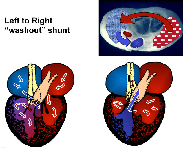
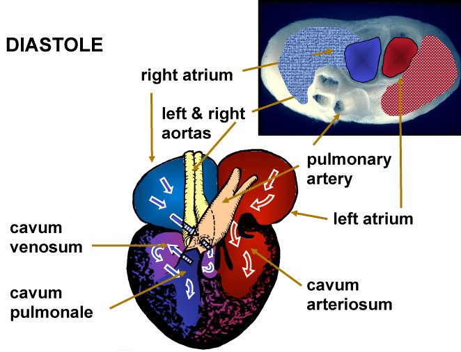
By contrast, turtle hearts are three-chambered and lack significant coronary circulation. Similar to fish hearts, turtle hearts are composed primarily of spongy myocardium that receives direct perfusion from the blood within. In addition, turtle hearts lack a complete septum between the left and the right ventricle. This contrasts with humans in which, the left and right ventricles are completely separated. Thus, turtle hearts can support both left-to-right shunts to better perfuse the body during exercise, as well as a right-to-left shunt to increase digestion and gastric acid secretion.

Therefore, “turtle CPR” focuses more on oxygenation. For those that ever have to resuscitate turtles, some pearls are:

  1. Turtle CPR is all about the airway. Small pieces of food can get stuck resulting in choking. In addition, turtles can indeed inhale water and drown – even in shallow water! This is despite the fact that turtles can oxygenate in water via cloacal (combined GI/GU tract) breathing…
  2. Get the turtle out of water
  3. Elevate the hind end of the turtle (to let gravity get rid of water)
  4. Straighten, then bend the front legs of the turtle. This may help squeeze out some more water from the lungs.
  5. Take the turtle to a vet afterwards! They will give oxygen (and usually antibiotics)

Also, everyone can benefit from a basic understanding of how to perform CPR. Dr. Glaucomflecken makes an excellent pitch here. And for those short on time to formally learn – remember to push hard and fast on the center of the chest (100-120/min, about the speed of Stayin’ Alive). Rescue breaths do not lead to better survival if you are not EMS trained. If you don’t believe that- even Walter White says so (don’t follow his example though -instead lock your elbows, use your core, and don’t hang out with psychotic drug lords)!

References

  1. https://cpr.heart.org/en/resources/cpr-facts-and-stats/out-of-hospital-chain-of-survival
  2. Jackson DC. How a Turtle’s Shell Helps It Survive Prolonged Anoxic Acidosis. News Physiol Sci. 2000 Aug;15:181-185. doi: 10.1152/physiologyonline.2000.15.4.181. PMID: 11390905.
  3. Cunningham LM, Mattu A, O’Connor RE, Brady WJ. Cardiopulmonary resuscitation for cardiac arrest: the importance of uninterrupted chest compressions in cardiac arrest resuscitation. Am J Emerg Med. 2012 Oct;30(8):1630-8. doi: 10.1016/j.ajem.2012.02.015. Epub 2012 May 23. PMID: 22633716.
  4. Farmer CG, Hicks JW. The Intracardiac Shunt as a Source of Myocardial Oxygen in a Turtle, Trachemys scripta. Integr Comp Biol. 2002 Apr;42(2):208-15. doi: 10.1093/icb/42.2.208. PMID: 21708712.
  5. Farmer CG. On the evolution of arterial vascular patterns of tetrapods. J Morphol. 2011 Nov;272(11):1325-41. doi: 10.1002/jmor.10986. Epub 2011 Jun 27. PMID: 21710654.
  6. https://farmer.biology.utah.edu/Hunt%20and%20Shunt.html
  7. https://crazycrittersinc.com/cpr-in-turtles-and-tortoises-yes-they-can-choke-and-drown/

How do painted turtles hibernate without oxygen for 3 months?

Short answer: Cutaneous respiration (breathing through their skin) and storing lactic acid in their shell.

Eastern Painted Turtle (Chrysemys picta picta).jpg
Eastern Painted Turtle (Chrysemys picta picta) Source: By Greg Schechter

While reading about green anoles and “hibernation” yesterday, I came across this intriguing line from Vitt and Caldwell’s Herpetology and felt compelled to read more:

Survival [of painted turtles during hibernation] is possible because of high tolerance for lactic acid buildup, which can be stored in the shell, and because their metabolic rate is reduced to 10–20% of their aerobic resting rate.

Painted turtles (Chrysemys picta) are the most abundant turtles in North America. There are four subspecies of the painted turtle, with the Western painted turtle being both the most colorful and common. Adult females are larger than adult males (10-25 cm vs 7-15 cm, weight 500g vs 300g). Painted turtles are named for their coloration and have red, orange, and yellow stripes found on their heads, necks, and tails. These turtles can live a long time – more than 40 years in the wild!

Painted turtles live in slow-moving freshwater. They bask for warmth on logs or rocks in the warmer seasons. In the winters, they hide and hibernate in the muddy bottoms of their freshwater habitats. Like many other reptiles, the sex of the hatchlings is determined by the temperature of the nest during the middle third of the incubation period (22-26 degrees C for males, >28 degrees C for females). Of course, global warming hurts these turtles’ chances for reproduction by causing more and more hatchlings to be female.

image
Metabolic rate depression during anoxic submergence of the painted turtle. Source

The hibernation of painted turtles is particularly interesting – they can survive in freezing waters (as low as 3 degrees Celsius) without oxygen for months at a time. Cold and anoxic environments allow the turtle to use a fraction of the energy of a similarly-sized aerobic mammal (e.g. < 0.01% of the ATP usage of a comparably sized rat). There are 2 possible limiting factors for the survival of a turtle facing such prolonged anoxia: 1) depletion of glycogen reserves and 2) buildup of lactic acid (a byproduct of anaerobic metabolism). As noted in the figure above, painted turtles can use as little as 0.01 kcal/kg/min in anoxic, cold environments. For a 500g turtle, this amounts to only 7.2 kcal/day. Painted turtles have high glycogen content in their livers, skeletal muscles, and heart – these stores are sufficient at this level of energy expenditure for the average turtle to last for 5.5 months! Clearly then, the major hurdle facing the anoxic turtle is the buildup of lactic acidosis.

FIGURE 3.
Postulated mechanism of shell buffering of lactic acid in painted turtles. Source:

Painted turtles can build up plasma concentrations of lactic acid as high as 200mM (by contrast, a normal level in humans ranges between 2-4 mM). The painted turtles counteract this phenomenon in several ways:

  1. Direct body fluid buffering – painted turtles have high baseline plasma bicarbonate concentrations (40mM), with periotneal and pericardial fluid having concentrations of 80-120 mM.
  2. The turtle’s shell. The shell may release calcium carbonate, allowing for further buffering. In addition, lactate may be directly sequesterd by the shell. The lactate can then be flushed out later in normoxic conditions. Even during hibernation, turtles can switch between a normoxic and anoxic states (which may explain turtles swimming below the ice of frozen ponds). Apparently, this mechanism is not unique to the painted turtle, but generalizes to vertebrate bone, the carpace of crustaceans, and the shells of snails. As further proof, soft-shelled turtles fare much poorer in anoxic waters, lending more credence to the shell mechanism.
Western painted turtle. Photograph by Clay Showalter.

For those interested in more reading, I’d recommend looking into the work of Professor Donald Jackson. He passed away in 2020, but he seems to have written much of the seminal work on painted turtle physiology during his time at Brown.

As a final note, adult painted turtles cannot survive truly freezing temperatures (-1 to -2 degrees C). However, hatchling painted turtles can via supercooling – which I may discuss this further in the future… Also, stay tuned for a discussion tomorrow comparing turtle and human CPR!

References

  1. Vitt, L. J., & Caldwell, J. P. (2014). Herpetology: An introductory biology of amphibians and reptiles. Pages 203-227.
  2. Taking the temperature of the painted turtle. Lab Anim (NY). 2013 Sep;42(9):315. doi: 10.1038/laban.376. PMID: 23965557.
  3. Jackson, D.C. Hibernating without oxygen: physiological adaptations of the painted turtle. J. Physiol. 543, 731–737 (2002).
  4. Jackson DC. How a Turtle’s Shell Helps It Survive Prolonged Anoxic Acidosis. News Physiol Sci. 2000 Aug;15:181-185. doi: 10.1152/physiologyonline.2000.15.4.181. PMID: 11390905.
  5. https://cob.silverchair-cdn.com/cob/content_public/journal/jeb/223/3/10.1242_jeb.222075/3/jeb222075.pdf

Why do green anoles (American chameleons) change color?

Short answer: It’s not entirely understood! Some factors inducing green-to-brown color change include increased light exposure, cooler temperatures, increased stress, and social interactions.

I was recently asked whether or not green anoles (Anolis carolinensis) change colors to brown when they “hibernate.” Green anoles are a tree-dwelling species of lizard native to the southeastern United States. They are known for being able to change colors between green and brown (see above). Pet stores sometimes refer to the green anole as the “American chameleon,” but this is a misnomer as it is not a true chameleon (which are Old World lizards). Not being from the southeastern United States, I haven’t seen many green anoles, so didn’t have a good prior on this question. In any case, doing some more reading, it seems green anoles don’t actually hibernate – instead they are just relatively inactive during the fall and winter.

It’s not fully understood why green anoles change colors. Some popular hypotheses for green-to-brown color change included as a response to increased light exposure, cooler temperatures, and increased stress. Additionally, some work suggests that brown coloration corresponds to subordinance, while green coloration indicates social dominance. During male-male interactions, the “winner” will usually be green while the “loser” will usually be brown. Interestingly, green anoles most likely do not change color to match their background. Field studies have actually observed that green anoles are mismatched to the surface they are sitting on more often than would be expected by chance. Although it’s still not entirely clear, I think the best answer to the question on green anole coloration during “hibernation” is that periods of inactivity and lower stress correspond to cooler temperatures, which in turn likely correlate with brown coloration.

By contrast, the how of green anole coloration is more clear. The green-to-brown transition occurs due to the stimulation of cells that contain melanin pigments (melanophores). Above these melanophores rest xanthophores (cells containing yellow pigment) and iridophores (cells containing “plates” that appear blue-green). Stimulation of melanophores (likely via hormonal mechanisms) causes upward migration of melanin, which blocks out light from xanthophores and iridophores. This results in the visible change of color from green to brown. Reaggregation of melanin within melanophores restores the original green color (Taylor et al.).

The green anole has spread to islands in the Pacific and Caribbean, where it is considered an invasive species. The changes in coloration make the green anole extra difficult to detect, requiring specialized field observers. In a recent paper, a group working in the Ogasawara Islands in Japan tried to combine remote sensing and machine learning to more efficiently detect these lizards. For those interested in more in-depth anole information, I recommend the Anole Annals run by Jonathan Losos from WUSTL.

Not really 100% related, but in my readings, I came across this fun figure from Herpetology with regards to dewlap color and head bobbing displays for male Anolis to attract females. After nodding my head for 6 hours straight in every ophthalmology interview – I certainly sympathize…

Figure 9.13 Three types of visual displays in Anolis lizards. Adapted from Echelle et al., 1971. from Herpetology.

References

  1. Vaughan GL. Photosensitivity in the skin of the lizard, Anolis carolinensis. Photochem Photobiol. 1987;46(1):109-114. doi:10.1111/j.1751-1097.1987.tb04743.x
  2. Yabuta S. and Suzuki-Watanabe A. 2011. Function of body coloration in green anoles (Anolis carolinensis) at the beginning of the breeding season: advertisement signaling and thermoregulation. Curr. Herpetol. 30(2): 155–158.
  3. Summers CH, Greenberg N. Somatic correlates of adrenergic activity during aggression in the lizard, Anolis carolinensis. Horm Behav. 1994;28(1):29-40. doi:10.1006/hbeh.1994.1003
  4. Jane F.F. Boyer and Lindsey Swierk. Rapid body color brightening is associated with exposure to a stressor in an Anolis lizard. Canadian Journal of Zoology95(3): 213-219. https://doi.org/10.1139/cjz-2016-0200
  5. https://www.anoleannals.org/2012/02/24/new-study-on-color-change-in-green-anoles/
  6. https://www.researchgate.net/publication/10584612_Sociality_stress_and_the_corpus_striatum_of_the_green_anolis_lizard
  7. Taylor, J.D., Hadley, M.E. Chromatophores and color change in the lizard, Anolis carolinensis . Z. Zellforsch. 104, 282–294 (1970). https://doi.org/10.1007/BF00309737
  8. Aota, T., Ashizawa, K., Mori, H. et al. Detection of Anolis carolinensis using drone images and a deep neural network: an effective tool for controlling invasive species. Biol Invasions 23, 1321–1327 (2021). https://doi-org.laneproxy.stanford.edu/10.1007/s10530-020-02434-y
  9. https://www.anoleannals.org/
  10. Vitt, L. J., & Caldwell, J. P. (2014). Herpetology: An introductory biology of amphibians and reptiles.
%d bloggers like this: